
상품상세 정보
안면근육 모형 1670
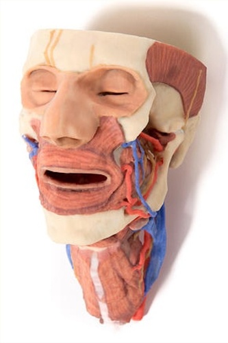
This 3D print focuses on the head and visceral column of the neck.
The face: On the head the right side is dissected to reveal the facial nerve and all its branches (temporal, zygomatic, buccal, marginal mandibular and cervical) which have been exposed due to removal of the parotid gland. The relations of structures embedded in the gland from superficial to deep (facial nerve, retromandibular vein, external carotid artery) are evident. Muscles of the head shown include temporalis, masseter and posterior belly of digastric. Arteries include facial artery, transverse facial artery and superficial temporal artery. The facial vein and transverse facial vein are clearly visible uniting to form the common facial vein which is joined by the retromandibular vein to form the external jugular vein. Viewed from the anterior aspect the face has been dissected to display some of the facial muscles around the mouth (buccinator [on the left], orbicularis oris and zygomaticus major) and the muscles of mastication (temporalis, masseter [right side only] and in the left infratemporal fossa the medial and lateral pterygoids are dissected. The lateral pterygoid is divided to allow the user to see the mandibular division of the trigeminal dividing into the lingual nerve and the inferior alveolar branch. The branches of the ophthalmic division of the trigeminal that supply the skin above the eyebrows and scalp (supraorbital [left only] and supratrochlear nerves [both sides]) are dissected. The submandibular gland is clearly visible below the mandible on both sides as are the facial arteries and veins as they course over the mandible.
The neck: The musculoskeletal portion of the neck have been removed to display the pharynx from behind, the larynx anteriorly and the neurovascular bundles laterally. The suprahyoid and infrahyoid muscles can bee seen on the neck. Indeed the vocal folds can be seen by looking up the length of the trachea from below. The cricothyroid muscle is also visible. The hypoglossal nerve can be seen winding around the lateral surface of the ECA and the external branch of superior laryngeal nerve is seen descending in the neck. The internal jugular vein, the common carotid artery and its bifurcation into ECA and ICA are clearly seen on both left and right. The vagus nerve in the carotid sheath is also visible. The superior thyroid artery branching from the ECA is seen descending in the anterior neck. The ansa cervicalis is visible emerging below the digastric muscle and descending on the surface of the internal jugular vein. The internal branch of the superior laryngeal nerve can be seen below the superior thyroid artery on the left. The internal branch of the superior laryngeal artery is visible on the left piercing the thyrohyoid membrane above the inferior constrictor where this muscle is attached to the hyoid bone.
Posterior view of the pharynx:The superior, middle and inferior constrictors are indicated on the pharynx wall. The oesophagus can be identified emerging from the lower end of the pharynx. The posterior horn of the hyoid bone acts as a useful landmark. The carotid sheath seen from behind clearly shows the vagus nerve and ist pharyngeal branches on the left. The recurrent laryngeal nerve is briefly visible on the left lying medial to the inferior thyroid artery. The occipital arteries are visible as they curve around the mastoid process. The vertebral arteries are seen either side of the brainstem as they enter the foramen magnum. The cerebellum has been removed to allow the fourth ventricle to be exposed. The cut surfaces of the cerebellar peduncles are clearly visible. A large portion of the posterior inferior cerebellar artery on the right is still visible as it winds around around the brainstem.
Cranial Cavity: The left and right orbits have been opened to reveal the orbital nerves and vessels along with the eyes and optic nerves. The optic chiasm, optic tracts and the lateral geniculate bodies are retained thus showing a large part of the visual pathways. The brainstem is cut at the level of the superior colliculi on the left and slightly lower on the right. The olfactory tracts and bulbs are also demonstrated. The origins of many of the cranial nerves from the brainstem are clearly visible.
| 상품명 | 안면근육 모형 1670 |
|---|---|
| 판매가 | 가격 전화문의 |
| 상품요약정보 | 독일제품. 두개골 안면근육 모형 |
| 국내·해외배송 | 국내배송 |
| 배송비 | 3,000원 (100,000원 이상 구매 시 무료) |
| 수량 |   |
결제 안내
배송 안내
- 배송 방법 : 택배
- 배송 지역 : 전국지역
- 배송 비용 : 3,000원
- 배송 기간 : 3일 ~ 5일
- 배송 안내 :