
상품상세 정보
흉추 - 골다공증 (A95)
Osteoporosis Model
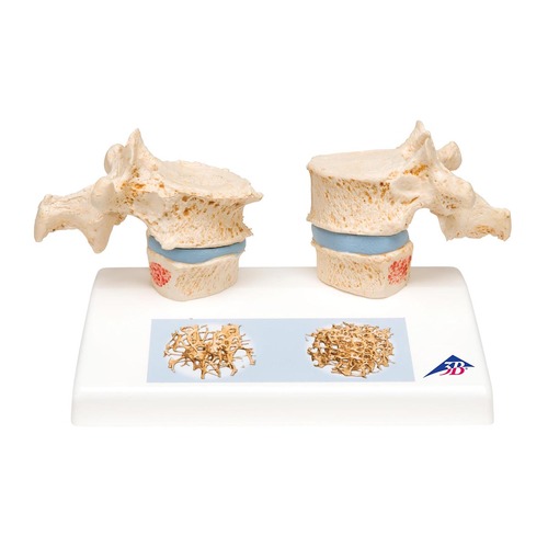
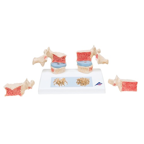
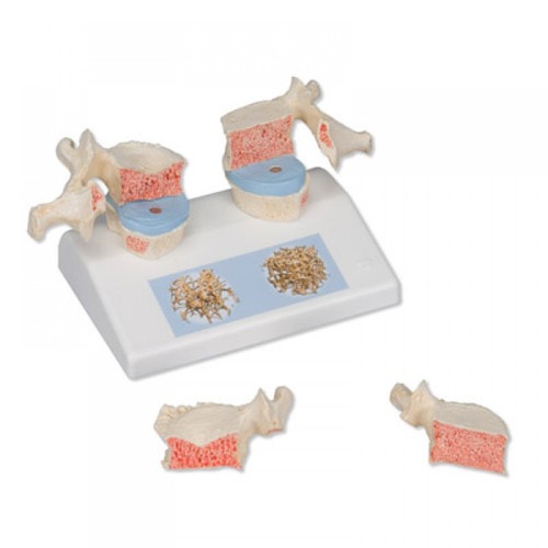
Impressive didactic model for comparing osteoporotic and normal thoracic vertebrae. Ideal for medical studies and patient consultation. The 11th and 12th thoracic vertebrae are shown.
Reproductions of sequential osteoporotic thoracic vertebrae with narrower intervertebral disc are located on the left of the stand. The upper vertebra is divided in the middle. The magnetically attached vertebral half can be removed easily to show the cut surfaces. This allows clear visualization of the fractured upper part of the vertebral body caused by sintering, i.e. collapse of the bony substance in the course and as a result of osteoporosis. Degenerative changes in the bone, manifested as osteophytes, are also identifiable.
For comparison, reproductions of two corresponding healthy vertebrae with intervertebral disc are provided on the right side. One half of the upper vertebral body is magnetically attached and can be removed.
A detail illustration on the base depicts two 3D micro CT images obtained from bone biopsies. These illustrate the microacrchitecture of the osteoporotic bone, which has a lower bone density compared to healthy bone.
| 상품명 | 흉추 - 골다공증 (A95) |
|---|---|
| 판매가 | 가격 전화문의 |
| 상품요약정보 | 독일제품, 골다공증이 표현된 11번 12번 흉추모형 |
| 국내·해외배송 | 국내배송 |
| 배송비 | 3,000원 (100,000원 이상 구매 시 무료) |
| 수량 |   |
결제 안내
배송 안내
- 배송 방법 : 택배
- 배송 지역 : 전국지역
- 배송 비용 : 3,000원
- 배송 기간 : 3일 ~ 5일
- 배송 안내 :